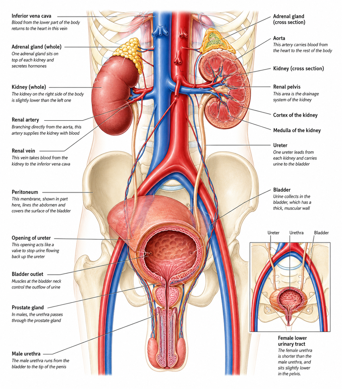
The urinary system consists of two kidneys, each linked by a ureter to the bladder, and a urethra, which connects the bladder to the outside of the body. The kidneys lie at the back of the abdomen, on either side of the spine. They are reddish-brown, bean-shaped organs, about 10–12.5 cm (4–5 in) long and 5–7.5 cm (2–3 in) wide. The ureters are thin, muscular tubes about 25–30 cm (10–12 in) long, and the bladder is a hollow, muscular organ located in the pelvis. The bladder’s lower opening is surrounded by muscle that helps to control the release of urine through the urethra.
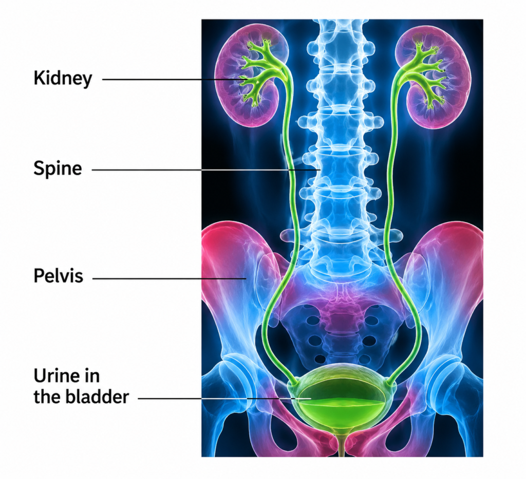
X-ray of urinary system
This specialized X-ray, known as an intravenous urogram (IVU), is used to highlight structures of the urinary system. The image clearly shows part of the kidneys; the ureters lying at either side of the spine; and the bladder. No abnormalities are seen.